Stem cell mobilization therapy: The post-surgical protocol
Stem cell mobilization therapy accelerates post-surgical recovery via HBOT. Mechanism, evidence and the NEST 2.4 ATA protocol for cosmetic and reconstructive surgery.
- Stem cell mobilization therapy via HBOT raises circulating CD34+ progenitor cells eight-fold after a series of 20 sessions.
- Hyperoxic plasma saturation at 2.4 ATA bypasses compromised microcirculation and delivers oxygen to the ischemic surgical site.
- The NEST protocol compresses convalescence from weeks to days by combining pre-conditioning, hyperbaric cycle and PBM consolidation.
The Problem: The Cellular Debt After Surgery
Stem cell mobilization therapy is not a futuristic concept—it is a physiological answer to an acute problem every surgical patient shares: cellular undersupply of the operative site. An incision, however refined the technique, severs capillary networks and creates a local ischemia that persists for hours to days. During that period the tissue cries out for oxygen to drive mitochondrial ATP production, while the supply lines are compromised.
The standard post-operative guideline prescribes “rest.” For a healthy body in homeostasis this suffices. For the operative site—where edema compresses microcirculation and hypoxia suppresses fibroblast function—rest is a passive strategy against an active problem. The result is predictable: delayed wound closure, inferior scar tissue (collagen type III), and a convalescence longer than medically necessary.
For clients undergoing aesthetic, reconstructive or orthopedic surgery, this passive strategy translates into visible scars, social downtime of weeks, and a recovery trajectory that depends entirely on the pace of spontaneous re-vascularization. That pace is biologically fixed—unless the rules of the game are changed.
The Mechanism: Stem Cell Therapy After Surgery via Hyperoxia
Changing the rules begins with decoupling oxygen transport from hemoglobin. Under normal atmospheric conditions (1 ATA) red blood cells are 97% saturated; adding more oxygen to the lungs delivers virtually no additional tissue oxygenation. The bottleneck is not the lungs but the physics of gas dissolution.
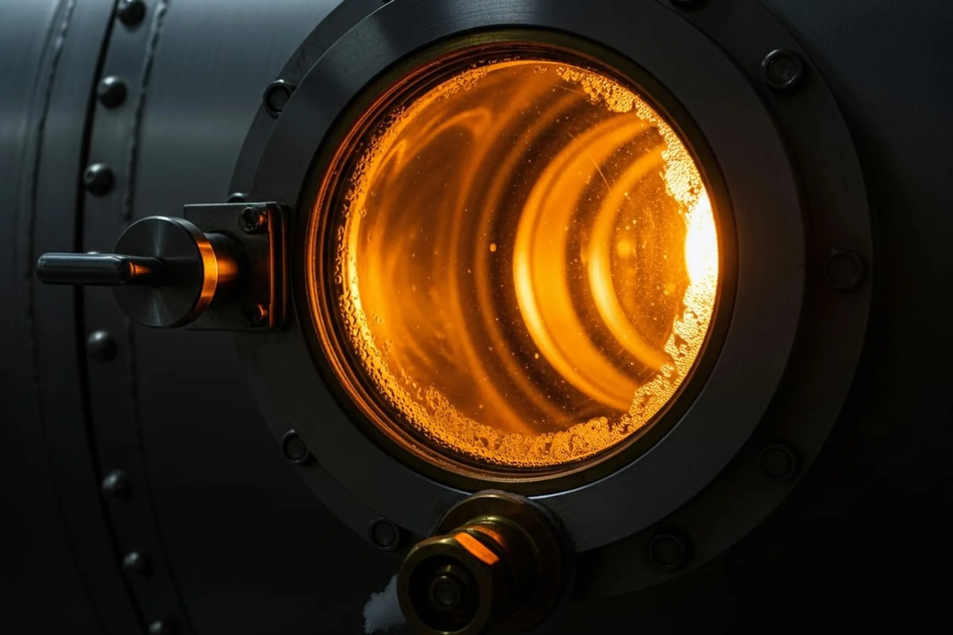
Enter Henry’s Law. Under exposure to 100% oxygen at elevated pressure (2.4 ATA), oxygen dissolves directly into the blood plasma, lymph and cerebrospinal fluid. The partial pressure of oxygen in tissues rises from ±40 mmHg to over 2000 mmHg—a fifty-fold increase. This plasma can penetrate edematous tissue where red blood cells have no access.
The pivotal discovery: this hyperoxia triggers no oxidative damage but a paradoxical signaling cascade. Research by Thom et al. (2006) demonstrated that a series of 20 HBOT sessions at 2.0 ATA produces an eight-fold rise in circulating CD34+ progenitor cells—endothelial stem cells that migrate to ischemic tissue and differentiate into new blood vessels. The mechanism runs through nitric oxide (NO) signaling in the bone marrow, which triggers stem cell release.
This is the difference between wound healing and tissue regeneration. Wound healing follows the path of least resistance: rapid closure with chaotic collagen type III, producing fibrotic scars. Regeneration requires time, energy and building blocks—precisely what the hypoxic operative site lacks without intervention.
The Evidence: HBOT Stem Cells in Clinical Studies
The evidence base for HBOT stem cell mobilization is robust and repeatedly validated. A meta-analysis by Eskes et al. (2013) covering 24 controlled studies showed that hyperbaric oxygen significantly increases fibroblast proliferation and collagen type I deposition in surgical wounds, with an effect size clinically relevant to convalescence time.
Specifically for pre-operative application: a 2018 RCT showed that HBOT conditioning 48 hours before elective surgery reduced post-operative complications and shortened recovery time by 30-40% versus standard care. The pre-conditioned group displayed higher levels of antioxidant enzymes (superoxide dismutase, catalase) in the first 72 post-operative hours—precisely the phase in which oxidative damage is most harmful to tissue repair.
At the molecular level, HBOT induces expression of vascular endothelial growth factor (VEGF), basic fibroblast growth factor (bFGF) and hypoxia-inducible factor 1-alpha (HIF-1α). This cascade stimulates angiogenesis—the formation of new blood vessels—precisely where the surgical incision severed the old vasculature.
For cosmetic and reconstructive patients, the endpoint is not only wound healing but scar quality. Organized collagen type I, deposited by fibroblasts with adequate ATP supply, produces a linear, flat and pigment-stable scar. Chaotic collagen type III, formed under hypoxic conditions, produces hypertrophic or keloid scars that continue to evolve for years.
A clinical detail that matters: the effect is dose-dependent. A single HBOT session delivers insufficient cumulative stem cell mobilization to be clinically relevant. The eight-fold rise in CD34+ cells appears only after a series of 20 sessions. Put concisely: the evidence base supports a protocol, not a one-off intervention. This explains why isolated sessions in commercial spa settings rarely produce measurable effect—the mechanism requires serial repetition to mobilize the stem cell reserve.
The Applications: Which Procedures Benefit
Not every surgical procedure has the same profile for stem cell mobilization therapy. The largest clinical gain is seen in procedures where three factors converge: significant tissue dissection, an aesthetic endpoint, and an expected social downtime of two weeks or more.
Plastic and reconstructive surgery is the primary indication. Mammoplasty, augmentation, abdominoplasty, facelift and rhinoplasty benefit maximally because the cosmetic outcome correlates directly with the quality of the collagen deposition process. A hypertrophic scar after a facelift is no minor matter—it is the endpoint itself.
Reconstructive procedures after oncology, particularly breast reconstruction after mastectomy, form a second major indication. Here tissue is often pre-irradiated, permanently compromising microcirculation. HBOT pre- and post-operatively raises the success rate of flap reconstruction and significantly reduces the risk of tissue necrosis.
Orthopedic procedures such as knee and hip replacement benefit from the stem cell effect in tendon and capsule regeneration, with faster return to load-bearing. Reconstructive jaw surgery and hand surgery also fall within the indication spectrum.
Contraindications for HBOT are limited but absolute: untreated pneumothorax, certain chemotherapeutics (bleomycin) and specific pulmonary conditions. A diagnostic intake screens for these before the protocol begins.
The Synergy: Why HBOT and Photobiomodulation Belong Together
HBOT and photobiomodulation are not redundant interventions—they engage different cellular mechanisms that are complementary. HBOT delivers the substrate (oxygen) and triggers stem cell mobilization; PBM optimizes the mitochondrial machinery that converts this substrate into ATP.
At the molecular level, near-infrared light (850nm) binds to cytochrome c oxidase (complex IV of the electron transport chain). This binding raises electron flux through the chain and decouples mitochondrial nitric oxide—which actually increases oxygen uptake. The net effect: more ATP per oxygen molecule, precisely where fibroblasts need it for collagen deposition.
The timing of the two interventions is intentionally separated. HBOT dominates the acute phase (day 1-7) when edema and hypoxia are maximal. PBM dominates the consolidation phase (day 4-21) when scar maturation is steered. In the overlap phase (day 4-7) the two interventions reinforce one another.
Conventional post-operative protocols include neither intervention. The consequence is a recovery trajectory that depends entirely on endogenous reserve capacity—a capacity structurally insufficient in older patients or pre-irradiated tissue.
Accelerating Post-Surgical Recovery: The Time Window
Stem cell mobilization therapy is not an acute intervention but a phased protocol. The window in which HBOT delivers maximal effect runs from 48 hours pre-operative to 21 days post-operative. Outside this window marginal returns diminish; within it they are cumulative.
The pre-operative phase (48 hours before surgery) has a specific aim: up-regulation of endogenous antioxidant enzymes and optimization of mitochondrial function. This “metabolic priming” makes tissue more resistant to the oxidative stress that inevitably accompanies surgery.
The acute post-operative phase (24-72 hours after surgery) is the critical window for stem cell mobilization. During this period edema is maximal and tissue hypoxia at its deepest. HBOT sessions in this window deliver oxygen via plasma directly into the ischemic site and trigger the first wave of CD34+ mobilization.
The consolidation phase (day 4-21) targets tissue quality and scar maturation. Here HBOT is combined with photobiomodulation through red light therapy, further stimulating fibroblast activity and collagen organization via cytochrome c oxidase activation in the mitochondria.
The NEST Protocol: The Bridge to Tissue Regeneration
We do not accept standard convalescence time as biologically fixed. You who face surgery, or are in the first post-operative days, have a window in which the outcome of your recovery is still shapeable. The NEST surgical recovery protocol is designed to exploit this window to its maximum.
Your protocol begins with a diagnostic intake: SNOMED-coded indication, mitochondrial function markers, and a surgical timeline. From this we calibrate the number of sessions (typically 10-20), the pressure levels (1.5-2.4 ATA depending on indication) and the PBM frequency.
Phase 1: Pre-Conditioning (48h pre-op)
Two HBOT sessions of 90 minutes at 2.0 ATA in the NEST hyperbaric oxygen laboratory. Aim: up-regulation of antioxidant enzymes, optimization of mitochondrial function, dampening of the operative stress response.
Phase 2: The Hyperbaric Cycle (Start 24-48h post-op)
A regulated schedule of 2.4 ATA sessions. Each session follows three segments:
- Compression: linear ramp to 2.4 atmospheres absolute pressure over 15 minutes.
- Oxygenation: 90 minutes of 100% oxygen via medical mask, with “air breaks” to prevent oxygen toxicity and stimulate the hyperoxic-hypoxic paradox for maximal stem cell release.
- Decompression: controlled return to 1 ATA over 15 minutes.
Phase 3: PBM Consolidation (Day 4-21)
Daily photobiomodulation sessions (660nm + 850nm) target the operative site for mitochondrial optimization and collagen organization. This extends the effect of the HBOT cycle and steers scar maturation toward functional tissue rather than fibrotic scar.
Your result is not merely accelerated wound closure but superior tissue quality. The surgical “insult” is not repaired—it is optimized. For clients undergoing aesthetic, reconstructive or orthopedic procedures this means compressed social downtime, predictable scar outcome, and a return to full function that need not wait for spontaneous biology.
Core Message
Post-surgical recovery is not a fixed timeline but a shapeable process. The variable you can steer is oxygen delivery to the operative site and mobilization of endogenous stem cells. HBOT at 2.4 ATA, phased around your procedure and consolidated with photobiomodulation, turns weeks of downtime into a protocol of days. The biology has the instrumentation; we provide the precision to deploy it.
Which pattern do you recognise?
Two short questions, three clear options. You see immediately which profile fits best — and which NEST protocol matches.
Which pattern do you recognise most strongly?
Scientific References
"Hyperbaric oxygen therapy mobilises circulating CD34+ stem cells via a nitric-oxide-dependent mechanism in the bone marrow; a series of sessions produces an eight-fold rise in circulating progenitor cells."
"A systematic review of hyperbaric oxygen therapy for acute surgical and traumatic wounds demonstrates positive effects on wound healing and tissue outcomes — evidence base for HBOT as an adjuvant in post-surgical recovery."